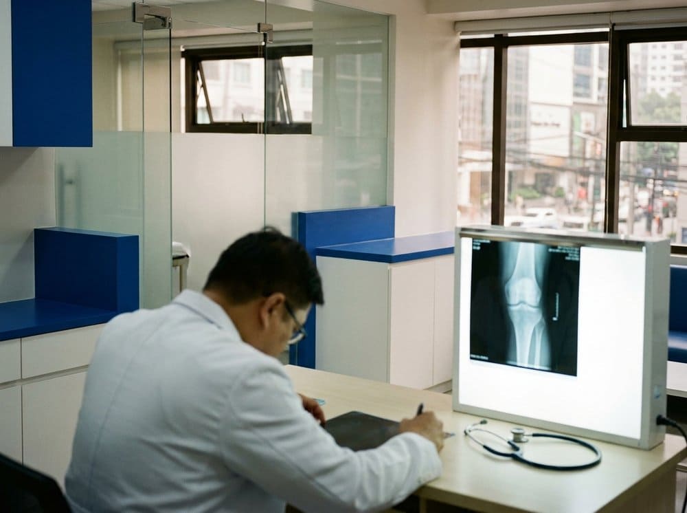
Most patients who walk into our Ayala clinic with knee pain have already had an X-ray taken somewhere. Sometimes it is a scanned film from a barangay health centre. Sometimes it is a crisp digital DICOM from Makati Med. Either way, the radiologist's one-line conclusion almost always ends with a roman numeral — "KL Grade III," or "KL II with early medial joint space narrowing." That numeral matters. It is the single most widely used grading system for knee osteoarthritis in the world, and it has been the international standard since it was proposed by British epidemiologists Jeffrey Kellgren and John Lawrence in their 1957 paper in the Annals of the Rheumatic Diseases.
But the grade is not a verdict. It is one data point. A Grade IV knee can feel surprisingly tolerable on a good day; a Grade I knee can hurt badly if the surrounding muscles are weak and the patient spends ten hours a day standing in an operating room or weaving through a jeepney commute. Knowing how to read the grade honestly is how you and your orthopedist pick the right next step.
What the X-ray is actually showing
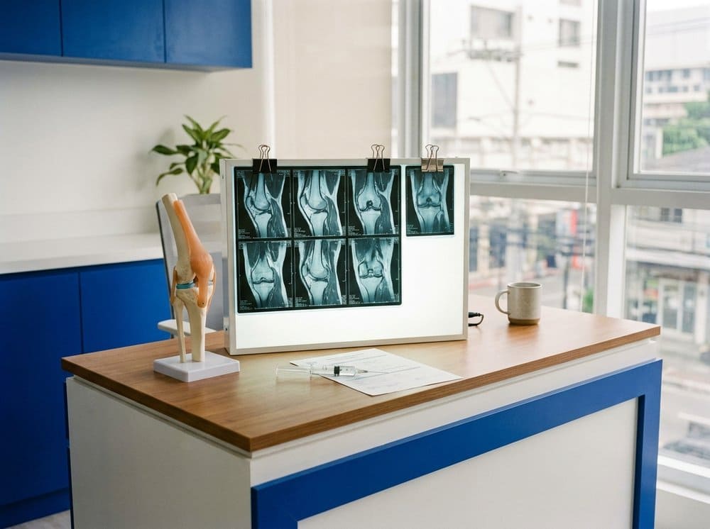
A standing (weight-bearing) antero-posterior and lateral view of the knee gives us four things:
- Joint space width. The gap between the femur and tibia. A healthy knee has roughly 4–5 mm of space, because cartilage is radiolucent and the bones do not actually touch.
- Osteophytes. Bone spurs that grow at the joint margins when cartilage wears down.
- Subchondral sclerosis. A bright, dense white line on the bone just beneath the cartilage surface — the body laying down extra bone where stress concentrates.
- Subchondral cysts. Fluid-filled pockets in the bone, visible as dark rounded holes.
Kellgren and Lawrence combined these four features into a 0–IV scale.
Grade 0 — No OA
A completely normal radiograph. No spurs, no narrowing, no sclerosis. If you have pain with a Grade 0 X-ray, the source is almost always soft tissue — meniscus, ligament, patellar tendon, or referred from the hip or lumbar spine. This is the moment when an MRI becomes useful, not a repeat X-ray.
Grade I — Doubtful
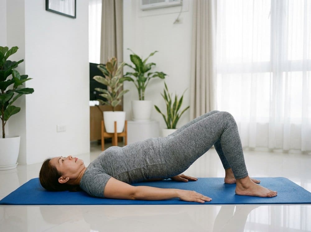
A tiny osteophyte, possibly at the tibial spine. Joint space still preserved. Radiologists sometimes disagree on whether this is OA at all. The clinical meaning: your cartilage is fine, but early changes are starting. This is the best grade to catch, because load management, weight control, and a structured home exercise programme — which we outline in our daily movement program for arthritic knees — can keep the disease essentially frozen for decades.
Grade II — Mild
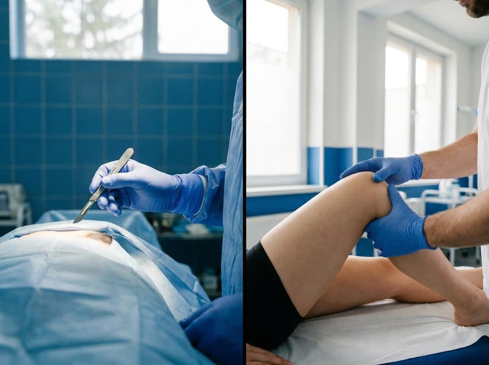
Definite small osteophytes. Joint space still looks largely preserved, perhaps with minimal narrowing. Patients at this stage typically describe stiffness after sitting through a long meeting, a deep ache after climbing the MRT steps, occasional swelling after a badminton game. First-line treatment is conservative: physiotherapy, activity modification, occasional NSAIDs, and — for patients whose symptoms outpace the radiographic findings — an intra-articular injection, usually corticosteroid for a flare or PRP for longer-term modulation.
Grade III — Moderate
Multiple osteophytes, definite narrowing of the joint space, some sclerosis, possibly minor deformity of the bone ends. This is the grade most of our injection patients fall into. The cartilage is thinning but still present, meaning biologic treatments can still help. This is also the grade where a structured 12-session osteoarthritis program pays off — we typically see WOMAC function scores improve by 30–40% in patients who complete it, even without surgery.
Grade IV — Severe
Large osteophytes, marked joint space narrowing (often bone-on-bone in the medial compartment), severe sclerosis, and definite deformity. This is the grade where we begin the honest conversation about total knee replacement — not because injections stop working entirely, but because their effect window shrinks. Even so, many Grade IV patients choose to delay surgery with a combination of viscosupplementation, focused rehabilitation, and preoperative conditioning that makes any eventual surgery easier to recover from.
"The radiologic progression of the disease does not always march in step with the patient's experience of it."
— Kellgren & Lawrence, Radiological Assessment of Osteo-Arthrosis, Ann Rheum Dis, 1957
Why two patients with the same grade can feel completely different
In our practice we routinely see pairs of patients with identical Grade III films — one is barely inconvenienced, the other cannot walk the length of Ayala Avenue. The discriminating factors are almost always the same:
- Quadriceps strength (the single strongest predictor of symptomatic knee OA in longitudinal studies)
- Body mass index, because every kilogram of body weight translates to roughly four kilograms of load through the knee joint during walking
- Alignment — a varus ("bow-legged") knee channels load onto the already-worn medial compartment
- Synovitis, which X-ray cannot show but MRI and ultrasound can
This is why our standard first visit includes not only the X-ray but a functional assessment, a WOMAC questionnaire, and a gait observation. The grade tells us what the bone is doing. The rest tells us what to do about it.
What to ask your orthopedist
If you are sitting in a consultation with your film on the lightbox, three questions will get you most of what you need: What grade am I, and which compartment is worst? Is my pain proportional to the grade, or is something else contributing? And given that grade and that pain, what is the most conservative thing we can try first? A good orthopedist will have clear answers — and will not reach for a surgical solution before the conservative options have been honestly tested.